Electron collisions, excitation and discrete spectra
By UK A Level Physics · independent study · myAudience
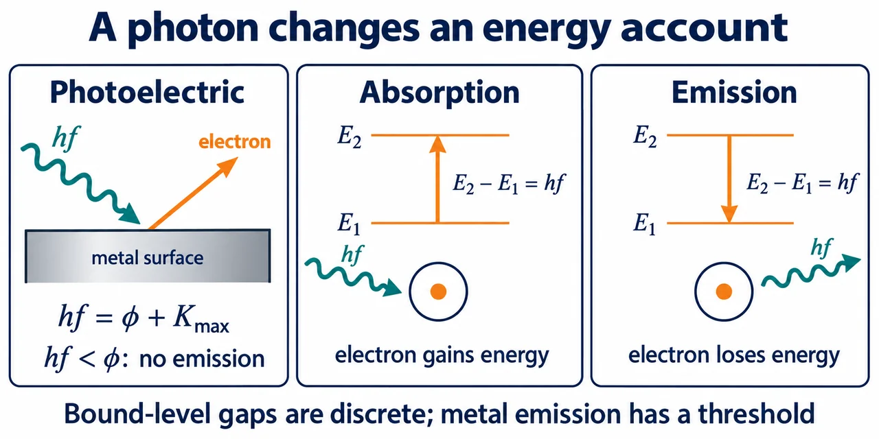
Image: illustration, not a documentary photograph. Original teaching model created for myAudience. · Original generated teaching illustration; no board endorsement or source textbook artwork reproduced.
Electron collisions, excitation and discrete spectra
Colliding electrons can transfer energy to atoms, but excitation and ionisation are different outcomes. This lesson assumes photon energy and eV–J conversion. Excitation raises a bound electron to an allowed higher energy level. Ionisation removes it from the atom, leaving an ion and a free electron. A collision must supply enough energy for the relevant change, and excess energy can remain as kinetic energy after ionisation. The incident electron does not need to be permanently captured to excite an atom.
The electronvolt is the energy change corresponding to one elementary charge moving through a potential difference of one volt: 1 eV = 1.602176634 × 10⁻¹⁹ J. It is not a unit of voltage. An electron accelerated through a potential difference of magnitude V gains eV joules under ideal energy-transfer assumptions, or numerically V electronvolts. Use this connection to compare collision energy with a supplied excitation gap or ionisation threshold; do not assume any arbitrarily small energy can create a bound excited state.
A fluorescent tube uses accelerated electrons in a low-pressure gas to excite and ionise atoms, sustaining a discharge. As excited atoms return to lower levels, they emit characteristic photons, commonly including ultraviolet radiation. A phosphor coating absorbs that radiation and emits visible light through its own energy processes. This is a conceptual account: real lamp efficiencies, gas compositions and coating chemistry are not inferred from the simple atomic level diagram, and mains apparatus is not a learner experiment here.
A line spectrum is evidence that the emitting atoms have discrete energy differences. For an emission from upper level E₁ to lower level E₂, hf = E₁ − E₂, a positive difference. Bound-state energies may be negative relative to the separated atom and electron; subtracting two negative numbers must still give a positive emitted energy for a downward transition. Absorption reverses the process when a photon matches an allowed gap. A spectrum measures differences, not directly the absolute position of every level.
Hydrogen spectra are a useful real example of discrete lines, but the worked example uses explicitly stipulated levels rather than copying a textbook diagram. A teacher-led observation through a diffraction grating can separate spectral lines; identifying wavelength from angle requires later wave reasoning and calibration. One gap does not imply only one line in the whole spectrum: different permitted transitions can have different gaps. Record which transition produced an answer and keep unit conversion visible so a negative-energy sign or nanometre conversion cannot silently reverse the physical interpretation.
The plate names the lower level E₁ and upper level E₂, so its positive gap is E₂−E₁. The worked emission example instead calls the initial upper level E₁ and final lower E₂; its gap is E₁−E₂. These symbols are local definitions, not contradictory sign rules: subtract lower energy from upper energy.
In the metal panel the incoming wavy arrow transfers a photon energy hf. The escaping electron arrow leaves the surface. The work function φ is the minimum energy to remove an electron from the metal; any remainder can be kinetic energy. The equation labels K_max because electrons can emerge with less energy after internal losses. The other panels show a bound atom. An upward electron-level arrow requires absorption; a downward transition emits a photon. Photon energy is the positive gap E₂−E₁ in either direction. The arrow describes a change of state, not a little electron flying vertically in real space. An atomic level resonance and a metal work-function threshold are different mechanisms.
**Original synthetic photoelectric table:** f/(10¹⁴ Hz) = 5.0,6.0,7.0; K_max/eV = 0.170,0.584,0.998. Using h = 4.14×10⁻¹⁵ eV s for this exercise, each extra 10¹⁴ Hz supplies 0.414 eV. Thus the gradient is 4.14×10⁻¹⁵ eV s and φ = 2.070−0.170 = 1.900 eV. Threshold f₀ = φ/h = 4.589×10¹⁴ Hz. The central data are constructed to lie exactly on this teaching line; that is not evidence of experimental precision.
**Demanding check.** Suppose each synthetic K value has an illustrative bound ±0.030 eV and f is exact. The end-pair limiting gradients are (0.968−0.200)/(2×10¹⁴) = 3.84×10⁻¹⁵ and (1.028−0.140)/(2×10¹⁴) = 4.44×10⁻¹⁵ eV s; the middle interval also contains both lines. Explain why a fitted slope near h supports the single-photon model but cannot by itself exclude a shared energy calibration error. At fixed f = 4.0×10¹⁴ Hz, doubling intensity leaves hf = 1.656 eV below φ and produces no emission in the elementary single-photon model. Above threshold it can increase the emission rate, not K_max. No stopping-potential measurement has been performed.
**Atomic transfer.** Supplied hypothetical bound energies are −5.0, −3.0 and −1.0 eV, with ionisation at 0. An atom initially at −5.0 eV can absorb a 2.0 eV photon to reach −3.0 eV, but a 3.0 eV photon matches no stated gap and is below ionisation. Assume isolated narrow bound levels and the elementary single-photon model; do not claim this excludes every broadening or multiphoton mechanism. From −1.0 eV direct decay to −5.0 eV gives a 4.0 eV photon; a cascade through −3.0 eV gives two 2.0 eV photons. With hc = 1240 eV nm, their wavelengths are 310 and 620 nm respectively. A 3.0 eV colliding electron can instead excite the 2.0 eV gap and retain 1.0 eV; an absorbed photon cannot leave a leftover fragment of itself. This distinction explains why collision excitation and resonant absorption need different accounts.
Worked example: A model atom has levels −5.0 eV and −2.0 eV. Find the wavelength for emission from the upper to the lower level using e = 1.60 × 10⁻¹⁹ C, h = 6.63 × 10⁻³⁴ J s and c = 3.00 × 10⁸ m s⁻¹.
The upper level is −2.0 eV, so ΔE = −2.0 − (−5.0) = 3.0 eV.
ΔE = 4.8 × 10⁻¹⁹ J.
λ = hc/ΔE = 4.14375 × 10⁻⁷ m.
Answer: About 4.1 × 10⁻⁷ m, or 410 nm to two significant figures.
Check your understanding: In a ground-state model atom, an allowed excitation gap is 4.0 eV and the ionisation threshold is 6.0 eV. Can a 3.0 eV collision cause either?
Answer: Neither of those changes.
Why: The supplied collision energy is below both specified requirements. Other possible levels must not be invented.