Pumping and distributing blood
By UK GCSE Biology · independent study · myAudience
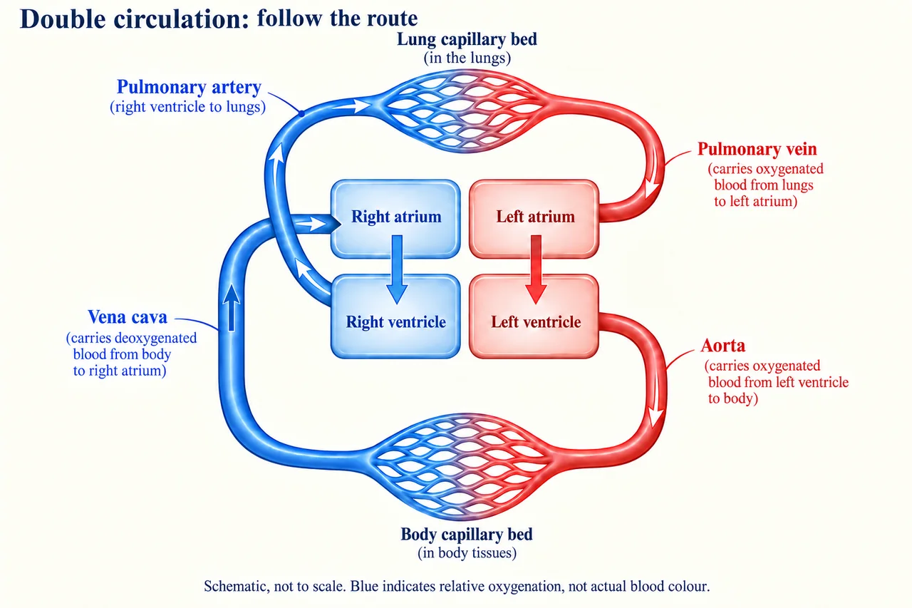
Original myAudience generated scientific illustration; not a documentary photograph or measured specimen. · Original generated project artwork; no source textbook figures reused. Sources support concept review, not copied-art licensing.
Pumping and distributing blood
The heart has four chambers and separates blood traveling to the lungs from blood traveling to the rest of the body. The right side pumps toward the lungs; the left side pumps to the body. Valves reduce backflow, and the left ventricle has a thick muscular wall for its pumping task.
Arteries carry blood away from the heart, veins toward it and capillaries through tissues. Their definitions concern direction, not oxygen content: pulmonary arteries carry relatively deoxygenated blood to the lungs. Capillary walls provide a short exchange route. Keeping direction and oxygenation separate prevents a common error when following blood through a heart diagram.
The vena cava returns blood to the right atrium; the pulmonary artery carries it toward the lungs. Pulmonary veins return it to the left atrium, and the aorta carries it toward the body. Arteries have muscular, elastic walls suited to pressure; veins commonly use valves to limit backward flow. At alveoli, a thin barrier, large surface and maintained air/blood supply support diffusion. Pacemaker cells in the right atrium coordinate the resting rhythm; a device can support some abnormal rhythms.
Worked example: Trace blood leaving the right ventricle before reaching the left atrium.
It enters the pulmonary artery
Gas exchange occurs in lung capillaries
Answer: It returns through pulmonary veins to the left atrium.
Check your understanding: Why are capillary walls thin?
Answer: They provide a short diffusion distance.
Why: Exchange becomes easier across a thin barrier between blood and surrounding tissues.