Communication through nerves
By UK GCSE Biology · independent study · myAudience
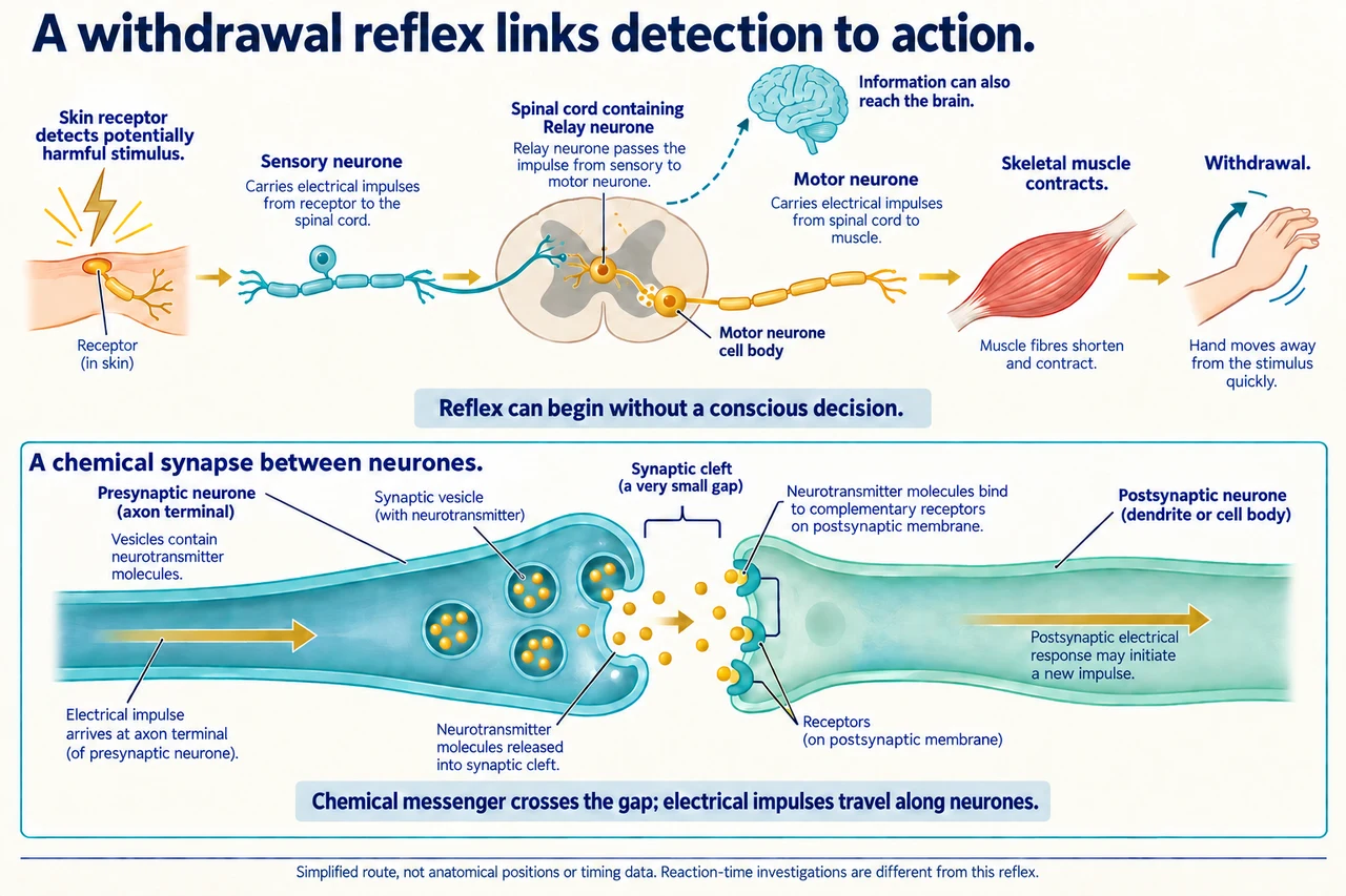
Original myAudience scientific illustration; not a documentary photograph or measured specimen. · Original generated project artwork; source concepts reviewed, no textbook figures copied.
Communication through nerves
Receptors detect stimuli; sensory neurones carry information towards the central nervous system, and motor neurones carry signals to effectors. At a chemical synapse, released neurotransmitter affects receptors on the next cell. An electrical signal does not leap unchanged across the gap. The pathway connects detection, communication and a response rather than making the receptor itself the responding muscle.
A withdrawal reflex commonly connects receptor, sensory neurone, relay neurone, motor neurone and skeletal-muscle effector through the spinal cord. It can begin without a conscious cortical decision, although information can also reach the brain. A voluntarily timed reaction is a different investigation: repeated observations and controlled conditions matter, and ruler-drop distance needs an appropriate relationship before it is reported as time.
Worked example: Trace a simple withdrawal reflex from a stimulus to muscle contraction.
A receptor activates a sensory neurone
Information passes through a relay pathway to a motor neurone
Answer: The muscle effector contracts.
Check your understanding: Why repeat measurements in a reaction-time investigation?
Answer: To assess variability and reduce the influence of an unusual trial.
Why: A single response can be affected by anticipation, distraction or measurement error.