How vision works
By UK GCSE Biology · independent study · myAudience
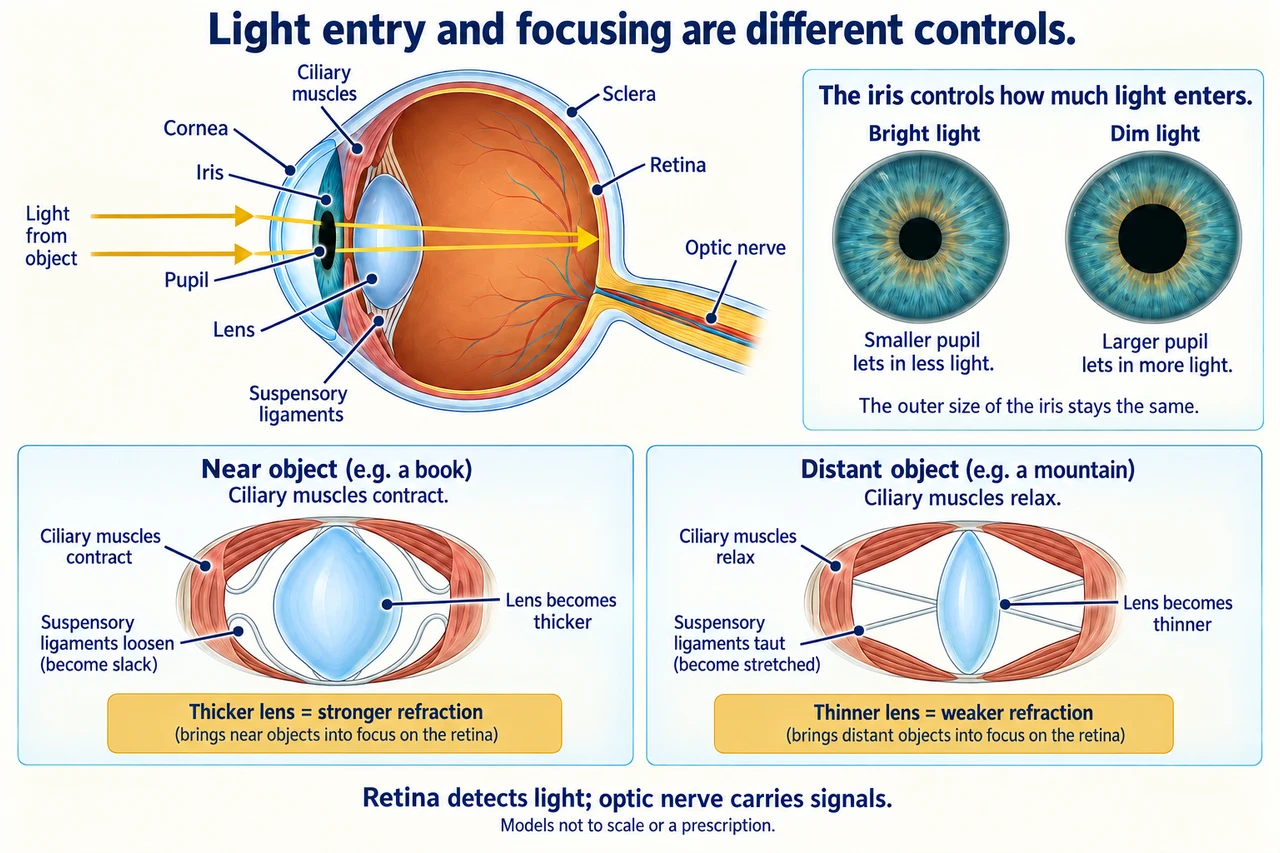
Original myAudience scientific illustration; not a documentary photograph or measured specimen. · Original generated project artwork; source concepts reviewed, no textbook figures copied.
How vision works
The cornea and lens refract light toward the retina, where receptor cells detect it. The optic nerve carries information from the eye. The iris controls pupil size, affecting how much light enters; focusing by accommodation is a separate process.
Accommodation changes lens shape through the ciliary muscles and suspensory ligaments. For a near object, the lens becomes thicker and more strongly refracting; for a distant object it becomes thinner. Short-sightedness and long-sightedness involve different focusing errors and corrective lenses. A classroom model describes the mechanism, not a diagnosis or prescription for a person.
The sclera provides a tough outer coat; the transparent cornea refracts incoming light, and the optic nerve carries signals from retinal receptors. For distance viewing, ciliary muscles relax, suspensory ligaments become taut and the lens becomes thinner. In a long-sightedness model, light would focus behind the retina and a converging correction can help; in short-sightedness, a diverging correction addresses a focus in front. These ray models explain errors without prescribing a lens for an individual.
Worked example: Distinguish the responses to a dim room and to reading a nearby page.
Dim light calls for a larger pupil
A near page requires accommodation of the lens
Answer: Pupil adjustment controls light entry; accommodation controls focus.
Check your understanding: When focusing near, do the suspensory ligaments tighten?
Answer: No; they loosen.
Why: Ciliary-muscle contraction reduces ligament tension, allowing the lens to become thicker.