How hereditary molecules are organised
By UK GCSE Biology · independent study · myAudience
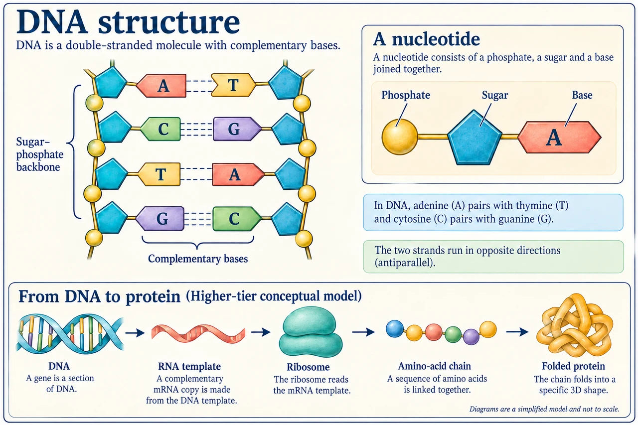
Original myAudience scientific illustration; not a documentary photograph or measured specimen. · Original generated project artwork; source concepts reviewed, no textbook figures copied.
How hereditary molecules are organised
DNA has a sugar–phosphate backbone and a sequence of bases in two strands. Each nucleotide contains sugar, phosphate and one base; complementary pairing links A with T and C with G across the strands. The sequence carries information, while the backbone provides the repeating structure. These are different aspects of the molecule.
Higher-tier extension: information is used through a template at ribosomes, arranging amino acids into a chain that folds into a functional protein. Coding variants can affect that chain or its shape; regulatory variants can affect when or how much a gene is used. Some changes have little observable effect. Connect DNA change, protein or expression change and cellular consequence rather than treating every mutation as automatically causing disease.
Worked example: In a DNA model, what pairs with A and with C?
Apply complementary DNA pairing
A pairs with T and C pairs with G
Answer: The complementary sequence is determined one base at a time.
Check your understanding: Must every mutation change a visible trait?
Answer: No.
Why: Some changes do not alter a protein’s function or have no detectable effect in that context.