Reading and scaling microscope observations
By UK GCSE Biology · independent study · myAudience
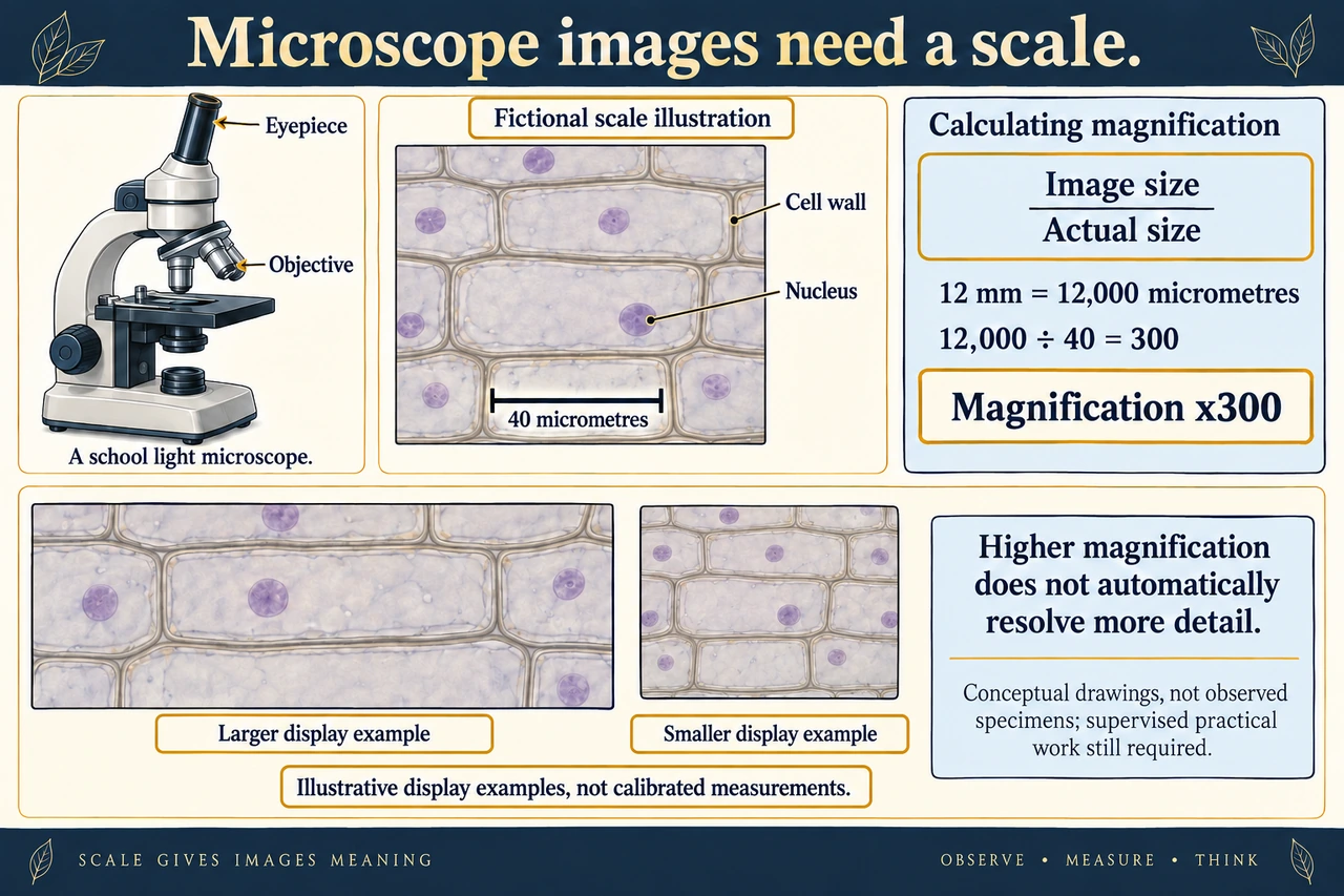
Original myAudience scientific illustration; not a documentary photograph or measured specimen. · Original generated project artwork; no textbook figure copied.
Reading and scaling microscope observations
A microscope image is evidence from a specimen; a drawing records the structures observed, not every organelle expected from a textbook. A clear biological drawing uses distinct lines and labels connected to the correct feature. Include a stated magnification or calibrated scale so a reader can relate the drawing to actual size. This lesson prepares reasoning for the practical study; it does not replace supervised microscope work.
Keep image size and actual size in matching units before calculating magnification. If an image measures 18 mm and the corresponding cell length is 30 μm, 18 mm is 18,000 μm and the magnification is 600. Total optical magnification in a simple school microscope is eyepiece magnification multiplied by objective magnification; increasing it does not automatically resolve more detail.
If a digital image is resized, its stated numerical magnification can become misleading, but an embedded scale bar resizes with the image. Distinguish what you directly saw from an interpretation about cell identity. Compare several cells and state the measurement direction rather than choosing the largest-looking cell as typical. Glass, stains and equipment require the teacher’s approved risk assessment and handling instructions.
Worked example: A 12 mm drawing represents a cell 40 μm long. Calculate its magnification.
Convert 12 mm to 12,000 μm.
Divide image length by actual length: 12,000 / 40.
Answer: The drawing is ×300; magnification has no length unit.
Check your understanding: Why is a scale bar useful after an image is resized?
Answer: It changes size with the image.
Why: The relationship between bar length and specimen length remains available when both are resized together.