Microscopes reveal different kinds of detail
By UK A Level Biology · independent study · myAudience
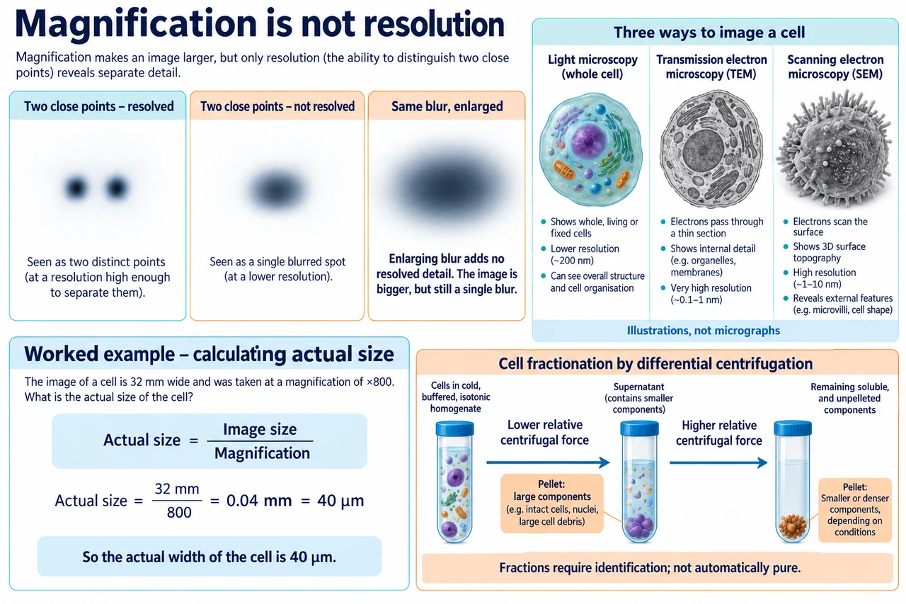
Original myAudience scientific illustration; not a documentary photograph or measured specimen. · Original generated project artwork; no source textbook figures reused. Sources support scientific review, not copied-art licensing.
Microscopes reveal different kinds of detail
Magnification makes an image larger; resolution determines whether nearby features can be distinguished. A larger blurry image does not necessarily provide more information. Electron microscopes can resolve finer detail than conventional light microscopes but involve different preparation and imaging constraints.
Cell fractionation separates components after cells are disrupted. Buffering, temperature control and centrifugation conditions affect what remains intact and where material collects. A fraction is evidence about composition, not an automatically pure sample of one organelle.
A transmission electron microscope can show internal features through a thin prepared sample; scanning electron imaging emphasizes surfaces. Preparation may create artefacts, and electron methods do not provide ordinary observations of a living hydrated cell. Fractionation uses cold, buffered and suitably isotonic conditions to limit degradation, pH disruption and osmotic damage. Successive centrifugation conditions separate larger or denser components earlier in a simplified model; the pellet still requires identification.
Worked example: A cell image is 32 mm long at ×800 magnification. Find its actual length.
Actual size = image size / magnification.
32 / 800 = 0.04 mm.
Convert millimetres to micrometres.
Answer: 40 µm.
Check your understanding: What limits whether two close structures appear separate?
Answer: Resolution.
Why: Magnification alone increases displayed size without necessarily separating adjacent detail.