The heart has a rhythm that can be adjusted
By UK A Level Biology · independent study · myAudience
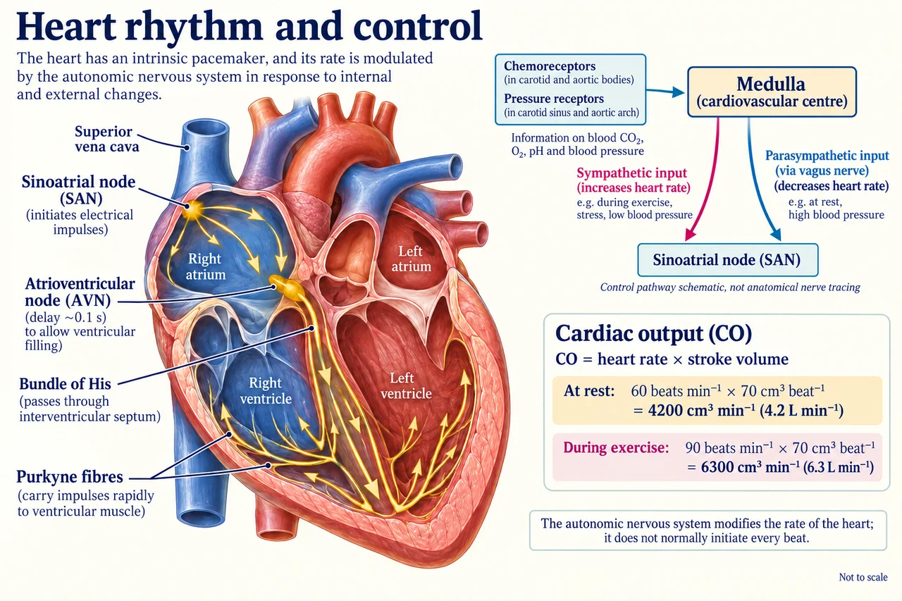
Original myAudience scientific illustration; not a documentary photograph or measured specimen. · Original generated project artwork; no source textbook figures or paragraphs reused. Sources support concept review, not copied-art licensing.
The heart has a rhythm that can be adjusted
The sinoatrial node initiates electrical activity in the heart. Activity spreads through the atria, then reaches the atrioventricular node, where a delay supports coordinated filling and ventricular contraction. Conducting tissue distributes activity through the ventricles.
Autonomic pathways modify the intrinsic rhythm. Receptors detecting pressure and blood chemistry contribute to feedback through cardiovascular control centres. A higher pulse is therefore not simply a voluntary instruction to each heart muscle cell; it emerges from a coordinated regulatory system.
The bundle of His and Purkyne fibres distribute activity so the ventricles contract in an effective sequence. Cardiac output is heart rate multiplied by stroke volume: an increase in one does not fix the overall change if the other also changes. In the worked example, a 50% heart-rate rise produces a 50% output rise only because stroke volume is held constant.
Worked example: Heart rate rises from 60 to 90 beats per minute while stroke volume stays at 70 cm³. Calculate the change in cardiac output.
Initial output = 60×70 = 4200 cm³/min.
New output = 90×70 = 6300 cm³/min.
Answer: An increase of 2100 cm³/min, or 2.1 dm³/min.
Check your understanding: Why is the atrioventricular delay useful?
Answer: It helps coordinate atrial and ventricular contraction.
Why: The timing supports filling before coordinated ventricular contraction.