Connecting observed organ structure with function
By UK A Level Biology · independent study · myAudience
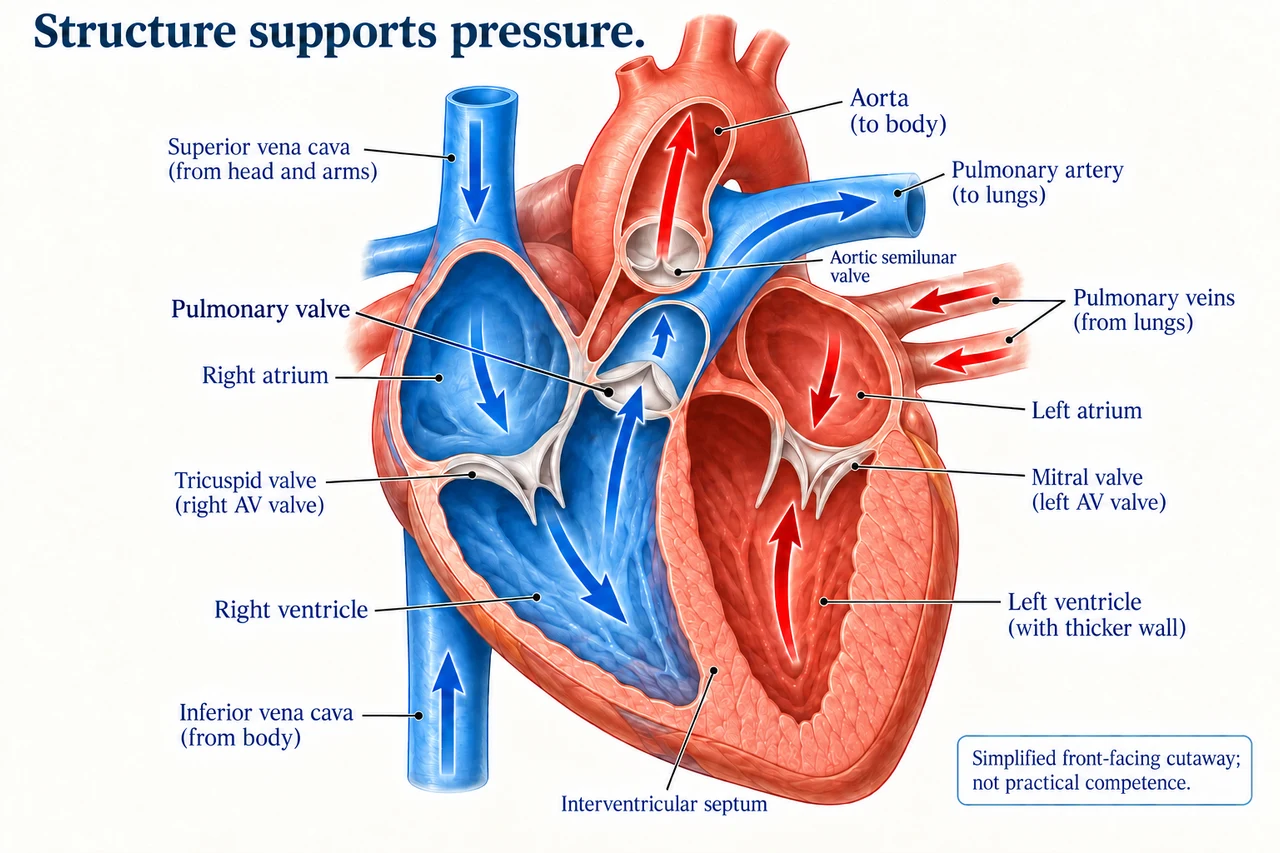
Original myAudience scientific concept illustration; not a photograph or measured specimen. · Original generated project artwork; no source textbook figures reused. Reference links support review, not a copied-art licence.
Connecting observed organ structure with function
A dissection or appropriate observation of a transport or exchange organ can connect three-dimensional structure with function. In a heart, chambers, valves and vessels are not interchangeable labels: follow the route of blood and the direction permitted by valves. An observation drawing should show what the specimen actually reveals, with a scale or dimensions where appropriate.
The thicker left-ventricular wall supports the greater pressure needed for systemic circulation, compared with the right ventricle serving the pulmonary circuit. Wall thickness and chamber size are different measurements. Orientation also matters: left and right refer to the organism, so a front-view image does not use the viewer’s left as the anatomical left.
This practical study permits suitable plant or animal exchange or transport structures; one heart diagram does not cover every option. Approved sourcing, respect for material and supervised risk assessment are part of actual work. This lesson teaches interpretation without cutting instructions. Viewing a model, image or simulation is not evidence that direct practical competencies have been observed.
Worked example: A labelled front-view diagram places the organism’s right atrium on the viewer’s left. Is that necessarily an error?
Anatomical labels refer to the organism’s own orientation.
Facing an organism reverses the viewer’s left/right relative to it.
Answer: No. Check the stated viewing direction and blood route before relabelling.
Check your understanding: Why is the left-ventricular wall usually thicker than the right?
Answer: It develops higher pressure for systemic circulation.
Why: The explanation links muscle structure with the pressure demands of the circuit, not with oxygen content alone.