Ultrasound: an echo measures a journey, not a diagnosis
By How things work · myAudience
myAudience original generated educational illustration · Original commissioned illustration
Ultrasound: an echo measures a journey, not a diagnosis
Diagnostic ultrasound turns emitted sound and returning echoes into spatial information. The transducer converts an electrical excitation into a mechanical wave and converts part of the returning disturbance into an electrical signal. NIBIB describes piezoelectric elements as a common implementation; not every possible transducer design must use the same ceramic. The established image-making mechanism is distinct from therapeutic ultrasound, which deliberately transfers energy to modify tissue. This lesson follows a diagnostic pulse-echo model without instructing anyone to scan a person or interpret an image clinically.
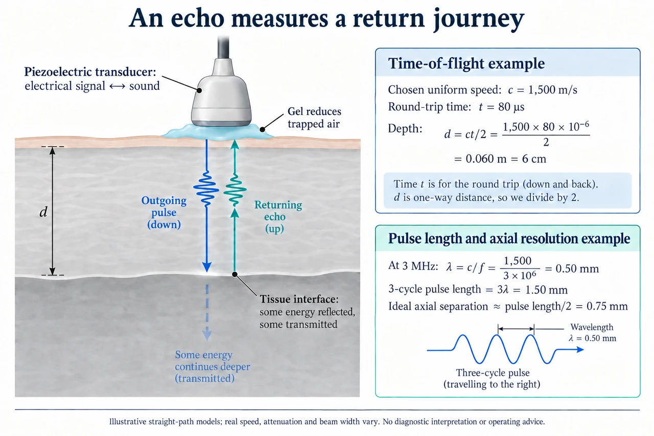
Follow the two arrows in the illustration. The outgoing pulse moves from the probe through coupling gel into tissue; part is reflected at an interface and returns to the probe, while part continues deeper. The gel reduces trapped air at the contact, which would interfere strongly with transmission. Reflection strength depends on the acoustic properties on each side and the angle of the interface; every boundary is not a perfect mirror. The simple flat layer is an original teaching model, not a picture of a named patient or an identified abnormality. Electrical-to-sound conversion does not generate X-rays or ionizing radiation, but it still introduces energy.
For a straight, uniform path with sound speed c and a pulse that returns after time t, the total distance travelled is ct. The interface is only half that distance from the probe, so d = ct/2. The plate chooses c = 1,500 m/s and t = 80 µs, where one microsecond is 10⁻⁶ s. Thus d = 1,500 × 80 × 10⁻⁶ / 2 = 0.060 m, or 6 cm. Using ct without dividing by two would mistakenly count outward and return travel as a one-way depth. The chosen speed is an assumption, not a measured average for every biological tissue.
A depth can be precise in arithmetic and still uncertain in inference. If the true straight-path speed were 1,600 m/s at the same measured time, the interface would be 6.4 cm deep. A reconstruction using 1,500 m/s would display 6.0 cm, underestimating that depth by 0.4 cm. Different tissue speeds, refraction, non-straight routes and repeated reflections can break the simple mapping. Echo amplitude also changes with attenuation and scattering; a weak or absent echo is not proof that a structure is absent. Multiple beam directions and appropriate reconstruction are needed for an image, rather than simply drawing a depth from one returned pulse.
Resolution has several directions and limitations. For the plate’s chosen 3 MHz pulse and 1,500 m/s speed, wavelength λ = c/f = 0.50 mm. A three-cycle pulse occupies a spatial length of 1.50 mm; in a simple ideal pulse-echo separation model, two reflectors along the beam need approximately half that pulse length, 0.75 mm, to produce distinguishable returns. This is an illustrative axial-separation estimate, not a clinical resolution rating. Lateral distinction depends on beam width and focusing, while out-of-plane slice thickness creates another limit. A smaller wavelength does not guarantee unlimited detail or depth: attenuation and signal quality matter, and shortening a pulse can trade signal energy against separation.
The frequency of the sound itself is not the same quantity as the repetition rate of emitted pulses or the frame rate of an image. A three-cycle 3 MHz packet lasts 3 / 3,000,000 = 1 µs in this model. Echo travel time may be much longer, and a scanner must distinguish returns belonging to different emissions. The MHz label therefore does not mean millions of complete tissue images per second. Doppler methods use shifts associated with motion and require their own geometry and processing assumptions; this single depth plate does not explain or validate blood-flow velocity measurements. Neither a pulse-echo image nor an attractive reconstructed colour field is an automatic diagnosis.
FDA explains that prudent professional use is important because acoustic energy can produce heating and mechanical effects. Absence of ionizing radiation is not a claim that any output, duration or untrained use is harmless. Appropriate exposure, medical need, equipment quality and trained interpretation belong to clinical practice. Equitable access also means communicating uncertain or unobtainable views honestly: device cost, training, patient comfort and body-specific imaging limitations can affect what evidence is obtained. New wearable or home imaging research must be evaluated for its actual intended use and study population; an experimental demonstration is not a universal replacement for clinical assessment.
Image: illustration, not a documentary photograph. The arrows show a partially reflected round trip through one uniform layer; the numerical depth and axial separation are chosen models. They are not a scan, patient measurement, calibration procedure, diagnostic threshold or universal resolution guarantee.
Worked example: A chosen straight-path echo returns in 100 µs through a uniform layer at c = 1,500 m/s. Calculate depth. A second hypothetical layer has c = 1,400 m/s at the same return time: compare true depth with the first reconstruction. For a separate two-cycle 2 MHz model at 1,500 m/s, estimate wavelength and ideal axial separation.
At the first assumed speed, d = 1,500 × 100 × 10⁻⁶ / 2 = 0.075 m = 7.5 cm.
At 1,400 m/s the depth is 0.070 m = 7.0 cm. Using the first speed would overestimate it by 0.5 cm; the echo time alone does not identify the correct speed.
λ = 1,500 / 2,000,000 = 0.00075 m = 0.75 mm. Two cycles occupy 1.50 mm; ideal axial separation is approximately 0.75 mm, separate from lateral resolution.
Answer: The depth models give 7.5 cm and 7.0 cm. The second pulse model gives 0.75 mm wavelength and approximately 0.75 mm ideal axial separation, without establishing clinical image quality.
Check your understanding: Why must the plate’s depth calculation halve ct? If its assumed sound speed is 1,500 m/s but the true speed is 1,600 m/s for the 80 µs echo, find the depth error. Does a 3 MHz sound frequency mean a 3 MHz image frame rate, or that ultrasound has no biological effects?
Answer: The echo makes an outward and return journey. Actual depth in the changed uniform model is 1,600 × 80 × 10⁻⁶ / 2 = 6.4 cm; the 6.0 cm reconstruction is 0.4 cm too shallow. Sound cycles, pulse emission and complete image acquisition are distinct rates. Ultrasound is non-ionizing but deposits acoustic energy and can have biological effects; neither calculation nor frequency establishes diagnosis or safe operation.
Why: Explain the measurement boundary and assumptions, calculate independently, and distinguish an illustrative signal from a validated clinical estimate.