Dialysis: transport, separation and what a model cannot promise
By How things work · myAudience
myAudience original generated educational illustration · Original commissioned illustration
Dialysis: transport, separation and what a model cannot promise
Dialysis uses selective transport to help replace part of lost kidney function. In haemodialysis, blood passes through a dialyzer outside the body and returns through a controlled extracorporeal circuit. A separate fluid, dialysate, receives selected substances across membranes. This is not whole blood poured into clean water, and the dialysate is not injected back as the returning blood. NIDDK emphasises that haemodialysis is not a cure for kidney failure and does not reproduce every kidney function. This lesson follows the established hollow-fibre exchange mechanism rather than instructing anyone to prepare or operate treatment equipment.
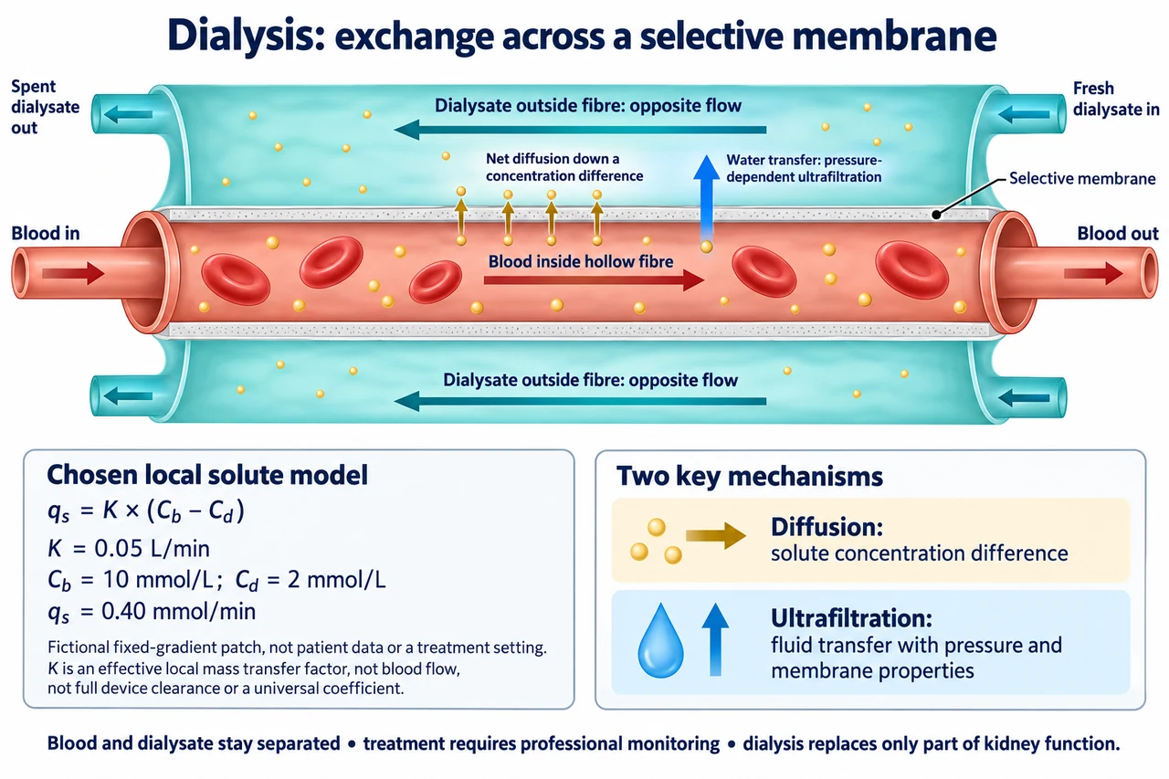
The main illustration flattens one hollow fibre so its two boundaries can be seen. Blood travels inside it from left to right; dialysate travels outside it in the opposite direction. The walls are selectively permeable membranes, with leader lines ending on the walls rather than on either bulk fluid. Small gold particles depict one selected permeating solute moving out because its local concentration is higher in the blood-side fluid. The large red cells remain on the blood side. Cell retention does not mean every dissolved protein or useful molecule is perfectly retained in every membrane: permeability, molecular properties and the selected treatment matter. Colours mark compartments, not oxygenated-versus-deoxygenated blood.
Diffusion is driven by a concentration difference for a solute that can cross the membrane. In a deliberately chosen local patch model, let q_s = K(C_b − C_d), where q_s is net solute transfer rate, K is an effective local mass-transfer factor and C_b and C_d are concentrations on the two sides. The image chooses K = 0.05 L/min, C_b = 10 mmol/L and C_d = 2 mmol/L. Thus q_s = 0.05 × 8 = 0.40 mmol/min. Units cancel to amount per time. K here is not the blood-flow rate, a universally measured membrane constant or a validated whole-device clearance. The fixed-gradient patch is an original two-fluid model, not patient concentrations or a treatment prescription.
The local model makes direction and limitation explicit. If C_d rose to 6 mmol/L while the other chosen conditions stayed constant, q_s would fall to 0.20 mmol/min. At equal 10 mmol/L concentrations, this ideal net diffusive rate would be zero, even though molecules continue moving in both directions. If C_d became 12 mmol/L, q_s would be −0.10 mmol/min: the sign represents net transfer into the blood-side fluid. It is therefore wrong to claim all substances always move out of blood. Clinical dialysate composition is carefully selected; identifying a substance as essential or waste does not allow the membrane to recognise moral categories or selectively remove it regardless of concentration and permeability.
Opposite flow helps sustain useful concentration differences along the exchange surface. It does not make the fluids mix or require every endpoint concentration to be equal. In a separate invented steady two-fluid example with negligible volume transfer and equal 1 L/min flows, the blood-side chosen solute concentration falls from 10 to 6 mmol/L, while dialysate enters at the blood-outlet end at 0 and leaves the opposite end at 4 mmol/L. The solute balance is 1×(10−6)=4 mmol/min lost on one side and 1×(4−0)=4 mmol/min gained on the other. At each paired end the blood-side concentration exceeds the local dialysate concentration by 6 mmol/L. These endpoints show a balanced possible countercurrent bookkeeping example, not a rating for the local patch coefficient or a blood-flow recommendation.
Removing water is a different transport question. In haemodialysis, pressure-dependent ultrafiltration transfers fluid across a permeable membrane, with membrane properties and opposing osmotic effects contributing to the actual relation. Diffusion of a dissolved waste alone does not specify water removal. Convection can also carry permeating solutes with fluid, so a complete mass balance may require both diffusive and convective terms. As pure unit practice, a fictional fluid-only model transferring 2 mL/min for 30 min would transfer 60 mL if its rate stayed constant; that arithmetic establishes neither a suitable clinical removal rate nor the resulting state of a person. The illustration separates the water arrow from the selected gold solute diffusion route.
An exchange calculation is only one part of a treatment system. NIDDK describes prescribed fluid composition and monitoring of blood flow and fluid removal. Membrane integrity, blood leaks, air, clotting, fluid quality and changes in the patient require professional controls and response. A pressure or temperature display alone cannot certify everything safe, and the conceptual fibre leaves out these engineered safeguards rather than implying they are unnecessary. Access, travel time, treatment burden, reliable electricity and safe water affect who can benefit. Responsible innovation evaluates safety and effectiveness in the intended context; portability or a newer membrane is not proof of a cure or of equitable access. Individual decisions remain with the person and their clinical team.
Image: illustration, not a documentary photograph. The separated hollow-fibre compartments and countercurrent arrows explain exchange; the chosen local equation explains direction and units without claiming a clinical clearance. Gold particles represent one selected solute, while the blue water arrow marks pressure-dependent transfer. No patient readings, membrane dimensions, needles, operating settings, fluid recipe or diagnostic threshold are supplied. Full membrane kinetics, patient-specific treatment adequacy and other dialysis modalities remain outside this mechanism lesson.
Worked example: In a chosen local two-fluid patch, K = 0.04 L/min, C_b = 9 mmol/L and C_d = 3 mmol/L. Calculate net solute transfer rate. Repeat with C_d = 8 and then 10 mmol/L, explaining the signs. For a separate steady bookkeeping example with equal 0.5 L/min flows and no volume transfer, the blood-side fluid falls from 8 to 5 mmol/L; dialysate enters at 1 mmol/L. Find its outlet concentration if solute mass is conserved.
q_s = 0.04 × (9 − 3) = 0.24 mmol/min. At C_d = 8, q_s = 0.04 mmol/min; at 10, q_s = −0.04 mmol/min, reversing the ideal net transfer direction.
The separate blood-side loss is 0.5×(8−5)=1.5 mmol/min. Dialysate must gain that amount, so 0.5×(C_out−1)=1.5 and C_out=4 mmol/L.
The countercurrent paired ends then differ by 8−4=4 mmol/L and 5−1=4 mmol/L. These balances do not apply the local K model to a whole device or establish treatment adequacy.
Answer: The chosen patch gives 0.24,0.04 and−0.04 mmol/min in the three cases. The unrelated conserved-flow example gives dialysate outlet 4 mmol/L. Neither determines patient treatment settings or complete kidney replacement.
Check your understanding: Does more solute diffusion automatically determine water removal, and do all useful molecules remain in blood? Why can net diffusion be zero without molecules becoming still? In the plate’s chosen patch, find q_s if K halves while concentrations remain 10 and 2 mmol/L, then identify what clinical conclusion cannot be drawn.
Answer: Water transport requires a separate fluid-transfer account, including pressure, membrane and osmotic effects; solute diffusion alone is insufficient. Permeability and concentration determine transport, so usefulness does not guarantee retention. Equal opposing molecular transfers can give zero net diffusion while microscopic motion continues. Halving K to 0.025 L/min gives 0.20 mmol/min in the chosen fixed-gradient model; it does not prove a device clearance, safe prescription, treatment adequacy or a cure.
Why: Explain the signal or transport boundary, identify model assumptions, calculate with units and distinguish a chosen model from clinical interpretation.